Holding a human heart and other surprises of anatomy lab, day 1
ListenTo many a bright-eyed first year medical student, there’s one experience that marks a real turning point or ‘rite of passage’ into the medical field. That moment when textbook studies leave the stage, and actual flesh and bone take the spotlight.
Enter gross anatomy 101. Human dissection.
Updated Jan. 1, 2015
To many a bright-eyed first year medical student, there’s one experience that marks a real turning point or ‘rite of passage’ into the medical field. That moment when textbook studies leave the stage, and actual flesh and bone take the spotlight.
Enter gross anatomy 101. Human dissection.
As an early hub for modern medicine and education, Philadelphia was a nexus for both the science and art of human anatomy, with famed anatomists like Dr. William Osler leading dissections in his University of Pennsylvania lab in the late 1800s, to renowned painters like Thomas Eakins capturing those cuts on canvas.
More than a century after these early pioneers made their first incisions, the anatomy lab still plays a vital, though evolving role in medical school. And it’s an experience that aspiring doctors like Frances Ding, Megha Mandalaywala and Samuel Werner – all first year medical students at Rowan University School of Osteopathic Medicine [RowanSOM] who are about to embark on their first lab of the year — may carry throughout their entire medical career.
The anticipation
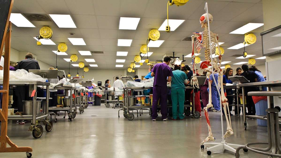
It’s a Friday afternoon in early November when several dozen first-year medical students at RowanSOM, most in their early twenties, filter into a lab room and prepare to meet the very first ‘patient’ of their formal medical training.
“I’ll be interested to see how emotional I’ll be,” said first-year Frances Ding. “She doesn’t know what kind of doctor she wants be, but says today will be an important test, whether she’ll be able to make it through the rest of her training, and more immediately, whether she can make it through the lab without fainting. “I think you get into medical school and you’re convinced you want to do it, but until you’ve had that experience, it’s hard to be 100 percent sure.”
For Megha Mandalaywala, a lot of buildup to this day came from family.
“My parents, when they found out when I was going to med school asked ‘when does anatomy lab start?'” recalls Mandalaywala. “Because they know that’s how you learn, the body is so fundamental to this career. That’s who we’re treating, we’re treating people and diseases that affect their bodies.”
Mandalaywala and Ding are nervous but also really excited.
“You have to understand, it’s a weird emotion to have,” says Mandalaywala, thinking about the fact that she and others will be spending the next three hours dissecting a dead body.
“It’s going to be intense,” says Samuel Werner, an aspiring primary care doctor, steps away from entering the lab, who describes his heart racing. “There’s an overwhelming presence that there are dozens of vessels that once carried souls within that room, no matter how [you] dismantle them [you] can’t hide what their original purpose was.”
Getting to know the ‘patient’
The lab has two sessions. Students in the alternating one have already made the first incisions, peeling back the skin and removing some of the ribs.
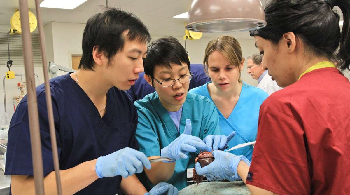
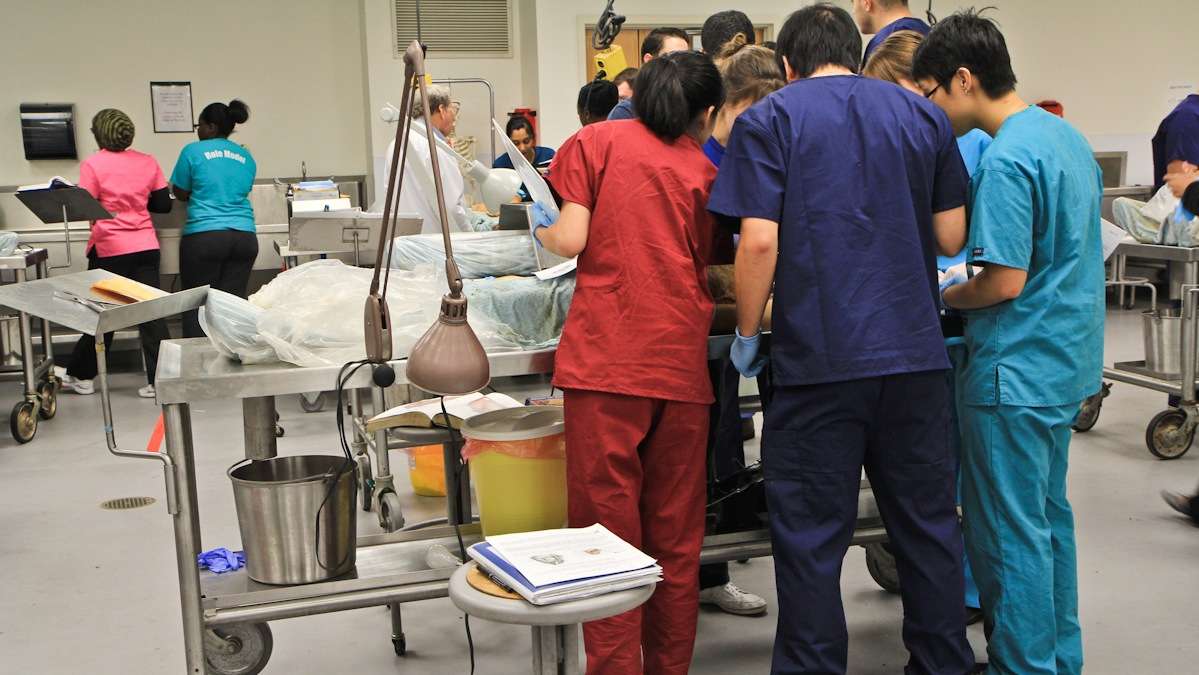
Werner, Mandalaywala and Ding, suit up in scrubs, break off into different groups, five per table. Today’s assignment? Removing and examining the heart.
“In text books everything has a very defined shape, that is not the case at all,” says Ding, as she and her group try to figure out how to remove the heart without damaging other vital nerves and muscles. “Everything is the shape it is to fit the body.”
15 bodies wrapped in cloth and plastic rest on metal tables. The heads and almost everything else are completely covered, except for the chest cavity, which is totally exposed, wide open, bleeding shades of bright burgundies and browns.
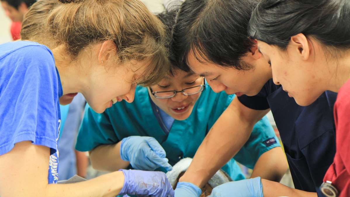
Call it adrenaline, youthful science lab excitement, or future doctor instincts kicking in, whatever apprehension Ding and others had going in has vanished. The room is abuzz with chatter as students debate how to make cuts without disrupting other important nerves and arteries, trying to identify all the nuanced features underneath the skin.
The only information students get about their bodies is the age, cause of death and the sex. That’s it. No names (though some students create names). Even so, Rocco Carsia, Rowan’s longtime anatomy teacher, says students will get to know them very well.
“The way we approach it here, we make the cadaver their first patient,” says Carsia. “Everything that goes along with it, the uncertainties, they learn secrets about the cadaver probably the patient themselves never realized. The kind of abnormalities they see, the students find them out in these investigations.”
In his more than 20 years, Carsia has only seen two students faint.
Changes to anatomy lab
Human anatomy education has changed in recent years, with more and more schools using three-dimensional imaging tools and other technological advances like ultrasounds. Even so, it’s an “adjunctional piece,” according to Jennifer McBride, head of histology at the Cleveland Clinic Lerner School of Medicine and leader of the International Anatomy Education Federation. “There really is no replacement for looking at a cadaver.”
In a 2009 survey of medical schools, McBride did find the number of hours allocated for anatomy lab have declined, as schools work to squeeze in more subjects. But “no school to my knowledge has gotten rid of the cadaver experience.”
McBride says schools are also experimenting with prosection verses dissection instructional techniques. In the case of prosection, students may zero in on a specific body structure prepped by an experienced anatomist.
Inside the RowanSOM lab, Carsia doesn’t lead any part of the class. Instead he walks around, checking in as students take charge. The point is for them to integrate what they learn here into their clinical development. Each group has a ‘lead dissector,’ who has previewed that day’s topic.
Werner and others focus on removing the heart, without disrupting other important nerves and parts of the body. It’s an art.
Each group’s body has a different story, a different history, and with that different dissection challenges. Mandalaywala’s table’s heart probably had a pacemaker. It has wires in it. The heart is also unusually large, requiring two hands to handle it.
“It’s really complicated,” she says, with a light laugh. “It’s a mess but we’re learning, right? That’s the whole process.”
Over the next two hours, students peel back layers, trying to identify and understand the different chambers and arteries.
“Until you actually see the way and feel the way its connected, understanding how one thing works in relation to another, you’re never going to have that special understanding, says Werner, pondering how to best maneuver cuts into the heart of a woman who died at the age of 107.
At another table, a student gasps, as she first lays eyes on the intricate tree branch-like patterns of the heart muscles.
As the 3 1/2 hour lab comes to a close, Mandalaywala and others provide in some ways the only real care they can to the bodies, redressing and wrapping them up. Spraying them with fluids to keep them from drying up.
It’s intimate, lifting hands, catching details and reminders of a life once lived.
Post lab processing
Stepping out of the lab, Mandalaywala is already looking forward to the next session.
“Finally, it feels like I’m in med school, you know?!” she says.
Reflecting afterwards, Ding found the experience validating.
“I’m surprised to see how easy I was able to disassociate the task of what I was doing from the person, the patient,” says Ding, adding that she’s grateful to those who chose to donate their bodies for the purpose of students’ learning and development. “I definitely think if I was faced in a real life situation, I wouldn’t be hung up on that.”
Werner is still processing, thinking about how the experience is changing his view of his own mortality and choices.
“I was literally holding in my hands a human heart. Not only a human heart, but a heart that belonged to a human” says Werner. “I could see reflected in my hands the life that she chose to lead. Once you hold that how can you not moving forward, enter into a new life where you won’t consciously be thinking about what choices you make will have on the physical reality around you?”
After the year-long course comes to a close this spring, the donated bodies are cremated, with the cremains returned to families. Students will hold a memorial service, where they’ll read essays, recite poetry, to honor those donated their bodies.
But for now, in what was just moments ago a booming room, full of life and energy, these bodies rest, wrapped up, waiting till next class, to serve as silent teachers for this next generation of doctors.
One year later
The Pulse caught up with Mandalaywala, Ding and Werner in December 2014, one year later, outside RowanSOM’s library, not the anatomy lab, in between exams.
All three successfully completed the course last spring.
“When you saw us, we were very starry eyed and very enthusiastic to get in,” said Ding, who said at times the lab was tough, spending hours upon hours examining the bodies. “It definitely was worth it now that I’m a second year.”
“That was a heck of an experience,” said Werner. “I can safely say looking back that I’m glad that the first day is behind me.”
Werner admits those first days and weeks in the anatomy lab were rough.
“I was just thinking, ‘I’m in the wrong place, medicine means seeing everything that could possibly go wrong with people,'” Werner recalled. “And it’s very easy to get in that mindset when you’re surrounded by cadavers.”
But as the class progressed, Werner recalled beginning to see the bodies in a new way – how they were meant to be and how it was “that one little aspect could lead to another” and in turn, have led to their decline in health.
“I could recognize as I looked around at the different bodies, ‘This one I can help. And this one, I can help too.'” said Werner. “‘Something must have gone wrong with this system, I can address that.'”
For Werner, the lab validated his pursuit of medicine, squashing any doubts he may have had.
“If there’s one thing this course has proven to me over the hours, weeks of time in the anatomy lab, even when there wasn’t an exam, just going in and studying the bodies…it showed me that it wasn’t a passing interest. It wasn’t something that I could check at the door.”
As for Ding, anatomy lab was an invaluable experience that has helped her understand other clinicals and concepts well beyond the lab.
“Even now as a second year I think back to my cadaver’s body that I spent a lot of time with and think about just different parts of his anatomy when I am learning about the pathology and all the different things that can go wrong.”
Mandalaywala agreed.
“By studying the visual part of anatomy with the cadavers, I was able to understand the relationships and how everything was laid out in our bodies so much better than when I was just reading it in a book. Because every body is different,” said Mandalaywala.
“It’s something we’ll always reflect back on.”
Looking ahead: what’s next, after anatomy?
Now in their second year, all three students agreed that their experience has intensified.
“If you thought first year was rough, let this be a warning to all the first years out there, it is nothing compared to second year,” said Werner.
“Life does suck right now,” says Mandalaywala, laughing, and acknowledging that she feels fortunate to even be in medical school.
“I’m definitely in survival mode right now,” said Ding.
So why is second year so hard?
“Volume of material. Volume of material. Volume of material,” said Werner, also laughing.
“We learned in the first year how the body is meant to work. We went through every system in the body for how one thing is supposed to relate to the other,” said Werner.
“In second year, we learn how those things can start to go wrong individually. And it’s something of a train wreck, which has an unfortunate, easy analogy to every single medical student’s mental state as they go through it.”
Mandalaywala and Ding laugh about it.
Mandalaywala explained that beyond delving into clinicals and pathology, students are also beginning to learn how to ‘doctor’ with patient actors and classes devoted specifically to bedside manner.
“The first one [patient scenario] we had, we were just like how do we begin? And it’s so much easier said than done.”
Looking ahead to 2015, Werner says his goal is to learn and absorb as much as possible from his clinicals and develop a deeper understanding of the human body, knowing what’s healthy and what’s sick.
Mandalaywala says she hopes to learn more about what happens when things go wrong in the body, and beyond that, “learning how to talk to patients will be exciting.”
Ding echoes that.
“A patient isn’t a multiple choice question,” she said. “When you get that comfort level, when you can go in and take a history…it doesn’t come to me naturally yet.”
“I don’t know when you go from the exam to ‘oh you passed’ to ‘oh I feel comfortable seeing a patient and diffusing whatever situation is behind those doors.’…So I’m hoping this next year that magically happens, so I feel ready for third year. So I don’t look a fool the first day,” said Ding, laughing.
“I heard that we do look like a fool the first day of rotations,” Mandalaywala responded, also laughing.
WHYY is your source for fact-based, in-depth journalism and information. As a nonprofit organization, we rely on financial support from readers like you. Please give today.
